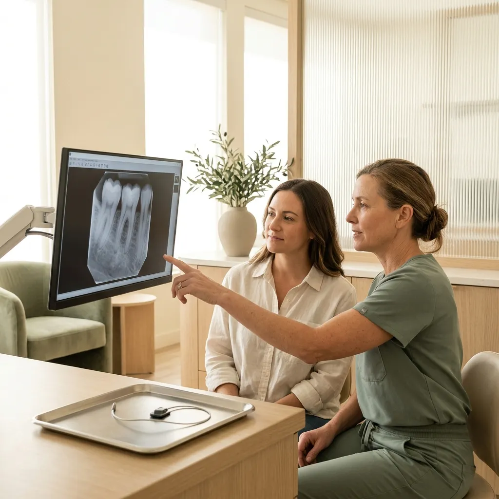
What it is
A digital X-ray uses a small electronic sensor instead of film. The image appears on the screen in a few seconds, can be enlarged and adjusted, and is stored in your file — no chemicals, no waiting.
The common types are bitewings (the back teeth from above and below, to find decay between them), periapicals (a whole tooth with its root, for pain or a root canal) and a panoramic image of both jaws in one picture.
Four digital bitewings: about one day of the background radiation everyone receives.
What it’s like
- We tell you which ones and why. X-rays are chosen for you, not on a fixed schedule.
- A small sensor. You bite gently on a holder for a second or two while the image is taken.
- On the screen at once. We look at the image together and explain what it shows.
What it changes — and what it doesn’t
What you gain
- Finds decay between teeth long before it hurts
- Shows roots, bone and infections around teeth
- Low dose, with results in seconds
- Images you can see and keep
Its limits
- A flat image: complex 3D questions may need a CBCT scan
- Early decay can be small, so some findings are watched and compared over time
How much radiation
A set of four digital bitewing X-rays gives an effective dose of around 0.005 mSv — about what you receive from natural background radiation in a single day in the US. A panoramic image is roughly two to three days of background radiation.
Since 2024 the American Dental Association no longer recommends lead aprons or thyroid collars for dental X-rays: modern digital sensors and a beam narrowed to the area being imaged protect you better, and a collar can block the very image we need. If you would still like one, just ask.